


VR – how lifesavers learn to save lives
At the University of Applied Sciences Vienna
Where VR helps the next generation of lifesavers save lives.
We’ve all seen them; those fly-on-the wall documentaries where sage and unassuming doctors perform life-changing operations with reassuring dexterity and confidence. We see the grateful patient, the relieved relatives and the methodical matter-of-fact way the whole complex business is carried out.
But there must have been a day when those same doctors were anything but confident; when they were trainees clutching nervously at their medical instruments, wondering just how much damage a slip could do. Would everything really happen the way it said in the textbooks?
Skilled and trusted
For most of us a level of responsibility that high would be near overwhelming, so just how do medical professionals get to be so confident, so highly skilled and trusted? The answer is increasingly medical VR, and while it may not be as glamorous as some Virtual Realities, it has quite likely saved more lives.
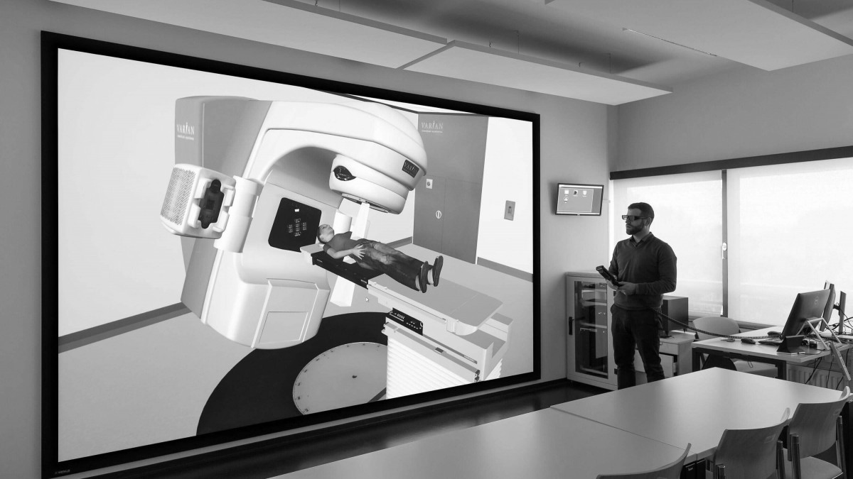
At the degree programme in Radiography at the FH Campus Wien (Campus of the University of Applied Sciences Vienna) VR now means that advanced radiotherapy treatments can now be taught under seemingly real conditions without risk.
Powerful and dangerous
And radiation therapy is an inherently risky business, using linear accelerators to deliver powerful radiation doses that have as much potential to do harm as good. The opportunity to practice in a realistic, risk-free environment is essential, and that’s exactly what Vienna system allows students to do.
Using controls that exactly match those they’ll use when qualified, and in an environment that mirrors real-world treatment rooms, they can now practice on a life-size, 3D, virtual patient, one that can display CT (computerised tomography) images, internal organs and show how different radiation doses are distributed through the body.
Precise imaging matters
Precision requires precision instruments, and that led to a Mirage WU12K-M projector supplied by Christie and active 3D shutter glasses supplied by Christie partner responsible for installation and project planning.
Sönke Dierking, Systems Team Manager at Virtalis takes up the story. “Christie Mirage WU12K-M was perfect here because we needed a WUXGA projector with high resolution and 3DLP technology for realistic 3D simulation. Using active shutter glasses meant perfect synchronisation with the projected image, the respective images for the left and right eyes refresh at 100 Hz and combined with back-projection any lag or shadowing is effectively eliminated.”
Spotting significant symptoms
That’s the thing about Medical simulations and the use of VR, it doesn’t necessarily have to be convincingly life-like but it must be a pin-sharp and accurate representation. Each patient and each case will present with unique symptoms, and a core skill of any clinician is to be able to recognise the commonalities and what they mean.
In the past, that skill could only be gained through hours of observing others at work, or by spending time with real patients and slowly building up a mental map of what to look for – only then could they distinguish significant symptoms from background diagnostic noise.
VR in medicine changes all that, and those students – who wondered if it really was going to look like it does in the textbooks – can now take their life-saving skills out into the real world knowing, quite literally, that they’ve seen it all before.